
More Information
Submitted: December 28, 2023 | Approved: January 09, 2024 | Published: January 10, 2024
How to cite this article: Karabinta Y, Karambé T, Konaté M, Sylla O, Coulibaly S, et al. Genital Condyloma in a 2-Year-Old Child Secondary to Circumcision: A Case Report. Ann Dermatol Res. 2024; 8: 001-003.
DOI: 10.29328/journal.adr.1001030
Copyright License: © 2024 Karabinta Y, et al. This is an open access article distributed under the Creative Commons Attribution License, which permits unrestricted use, distribution, and reproduction in any medium, provided the original work is properly cited.
Keywords: Accumulated condylomas; Circumcision; Child

Genital Condyloma in a 2-Year-Old Child Secondary to Circumcision: A Case Report
Karabinta Y1,2*, Karambé T3,4, Konaté M5, Sylla O1, Coulibaly S1, Gassama M1 and Dissa L1
1University Hospital Center of Dermatology, Bamako, Mali
2Faculty of Medicine and Dentistry, Bamako, Mali
3Dermatologie Teaching Hospital of Bamako, Mali
4Gabriel TOURE Teaching Hospital, Bamako, Mali
5Kati Teaching Hospital, Mali
*Address for Correspondence: Dr. Karabinta Yamoussa, Lecturer, FMOS/USTTB, University Hospital Center of Dermatology, Bamako, P.O Box 251, Mali, Email: [email protected]
Accumulated condylomas are exophytic tumors with a warty and hyperkeratosic surface due to the Human papillomavirus (HPV). Its prevalence in children is difficult to estimate due to limitations in epidemiological data. Its recurrent character is found in 30% of patients. Its management is very complex in children because of skin fragility. Circumcision is an operation consisting of the removal of part of the foreskin. This practice is done either with a simple knife or a pair of non-aseptic scissors which can be a source of contamination including HPV (Condyloma). Traditional circumcision does not seem to be reported in the literature as a mode of contamination. We report a case of genital condyloma in a child 2 years after circumcision. This is a 2-year-old male with no medical history but with a surgical history of circumcision that was brought by his parents in dermatological consultation for papular lesions accumulated on the penis. At the interrogation, we found the notion of recent circumcision performed by a tradithérapeute. The physical examination finds a good general condition. Dermatological examination reveals on the glans of multiple papules, exophitic, with warty and hyperkeratotic surface, of normal skin color. Furthermore, the physical examination of both parents was normal. The diagnosis of accumulated condyloma secondary to probable circumcision was retained before the clinical appearance of the lesions. Two electrocoagulation sessions spaced one month apart under local anesthesia were the treatment with a favorable evolution.
Accumulated condylomas are exophytic tumors with a warty and hyperkeratotic surface, ranging in color from erythematous to grayish or flesh-colored, caused by the Human Papilloma Virus (HPV) [1]. Their prevalence is steadily increasing, estimated at 1% in young adults, with an incidence of 107/100,000 inhabitants [2]. Estimating prevalence in children is challenging due to limited epidemiological data [2,3], and reported cases are on the rise [3].
Recurrence occurs in 30% of patients, making management complex in children due to skin fragility. Circumcision, the removal of part of the foreskin, is considered a rite of passage to adulthood in many African countries [4]. Traditional circumcision can lead to complications, performed with a simple knife or non-aseptic scissors, potentially causing contamination, including HPV (Condylomas). Among the modes of contamination of anogenital condyloma in children, traditional circumcision does not seem to be reported in the literature. We report a case of genital condyloma in a 2-year-old child.
This involves a 2-year-old male with no medical history but a surgical history of circumcision. The child was brought by his parents to the University Hospital Center of Dermatology on April 12, 2023, for accumulated papular lesions on the penis. Upon inquiry, a recent circumcision performed by a traditional healer was revealed. This circumcision resulted in a large erosion complicated by necrotic wounds. The parents sought treatment at a local community health center, where daily and twice-daily dressing changes, antibiotics (Erythromycin 250mg twice daily), and analgesics (Paracetamol Syrup three times daily) led to wound healing after several weeks. A few months later, small papules appeared on the penis, gradually increasing in number and size, prompting a consultation at the Dermatology Department of the University Hospital Center in Bamako. Clinical examination revealed the child in good general condition with well-colored conjunctiva, no pallor, signs of dehydration, or jaundice. The brachial circumference and growth curve were normal. Dermatological examination showed (Figures 1,2) multiple exophytic papules on the glans, with a warty and hyperkeratotic surface, and normal skin color, resembling a rooster's comb. Oral mucosa and appendages appeared normal. The remainder of the clinical examination, including both parents' dermatological assessment, was normal. The diagnosis of accumulated condyloma secondary to circumcision was established based on the clinical appearance. As part of the assessment, a retroviral serology (HIV) and complete blood count returned normal results. The patient underwent two sessions of electrocoagulation, one month apart, under local anesthesia. Electrocoagulation was combined with local care using antiseptic and Zinc Oxide ointment to promote re-epimerization. Therapeutic evolution was marked by the disappearance of lesions and the complete healing of erosions.
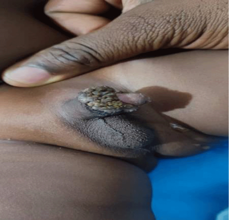
Figure 1: Multiple exophytic papules on the glans.
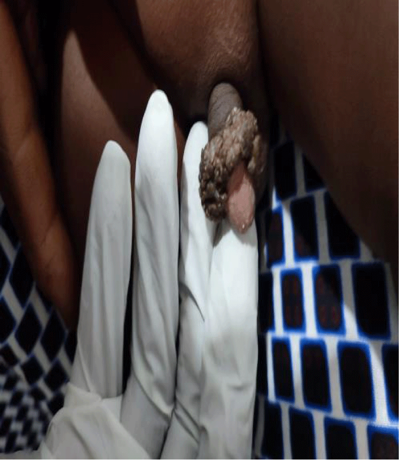
Figure 2: Papules with a verrucous and hyperkeratotic surface of normal skin color.
Genital condylomas are typically a disease of sexually active young adults. While sexual transmission is more considered in adults, identifying the mode of contamination remains problematic in children. Currently, three contamination modes are known in children: horizontal (auto or hero-inoculation), vertical (materno-fetal contamination), and sexual transmission. This clinical observation raises suspicion of the role of traditional circumcision. Traditional circumcision, as practiced in various African countries, generally involves a simple knife, without anesthesia, with the body covered in white clay, and the penis wrapped in leaves collected from nature [5]. This can be a source of contamination by various pathogens, including bacteria, fungi, and viruses such as human papillomavirus (HPV), due to the lack of aseptic practices, and the same knife may be used to cut the foreskin of several young boys. If this circumcision served as an entry point for HPV in a young child immunologically susceptible to infection due to his young age, we also considered the possibility that if one of the asymptomatic carrier parents provided care for the child, they could transmit the infection manually. Two authors, B. Dahmani, et al and E. Siegfried, et al, have suggested the concept of sexual abuse in a young girl [6]. Could we consider the manipulation of this little boy's penis by a woman such as housemaids and close relatives?
In the literature, several studies have mentioned the effectiveness of liquid nitrogen and electrocoagulation in children [7-9]. We chose electrocoagulation to shorten the duration of treatment because the lesions were accumulated, and cryotherapy would take too long to destroy these lesions.
The discovery of accumulated condyloma in a 2-year-old child on post-circumcision erosions raises a dual problem, namely the non-compliance with infection prevention measures and the improper practice of circumcision. It is essential to medicalize any circumcision procedure to avoid such complications.
Ethical consideration
The free and informed consent of both parents has been obtained regarding the image and publication.
- Mael-ainin M, Senouci K. Condylomes anaux de l'enfant [Anal warts of the child]. Pan Afr Med J. 2014 Jan 6;17:1. French. doi: 10.11604/pamj.2014.17.1.3736. PMID: 25184018; PMCID: PMC4149791.
- Collet-Villette AM, Gaudy-Marqueste C, Grob JJ, Richard MA. Prise en charge des condylomes anogénitaux profus de l'enfant par laser CO2 [Carbon dioxide laser therapy for anogenital warts in children]. Ann Dermatol Venereol. 2007 Nov;134(11):829-32. French. doi: 10.1016/s0151-9638(07)92825-2. PMID: 18033061.
- Lukasiewicz E, Aractingi S, Flahault A. Incidence and management of external anogenital warts in the general medicine department. Ann Dermatol Venereol. 2002; 129: 991-6.
- Cohen BA. Warts and children: can they be separated? JAAPA. 1999 Dec;12(12):63-8, 71-2, 75-6. PMID: 11010083.
- VOA Afrique. Circumcision, a dangerous rite of passage to adulthood. https://www.voaafrique.com/
- Dahmani B, Bouchennack K, Stamhouli OB. Anogenital condylomas in children; not to overlook sexual abuse. A case report. Ann Dermatol Venereol. 2016; 16(38): 301-3.
- Siegfried E, Rasnick-Conley J, Cook S, Leonardi C, Monteleone J. Human papillomavirus screening in pediatric victims of sexual abuse. Pediatrics. 1998 Jan;101(1 Pt 1):43-7. doi: 10.1542/peds.101.1.43. PMID: 9417149.
- Beutner KR, Ferenczy A. Therapeutic approaches to genital warts. Am J Med. 1997 May 5;102(5A):28-37. doi: 10.1016/s0002-9343(97)00181-2. PMID: 9217660.
- Marcoux D, Nadeau K, McCuaig C, Powell J, Oligny LL. Pediatric anogenital warts: a 7-year review of children referred to a tertiary-care hospital in Montreal, Canada. Pediatr Dermatol. 2006 May-Jun;23(3):199-207. doi: 10.1111/j.1525-1470.2006.00218.x. PMID: 16780463.